In STEM with Science and Technical Writing, taught by Dr. Crowthers, we work on both individual and group projects. A large part of the year is spent working on STEM I projects, which are independent research projects in a chosen STEM topic. Over the course of 6 months, we brainstorm potential topics, conduct extensive research for our projects, and test novel research ideas in science, engineering, or math topics to present in fairs.
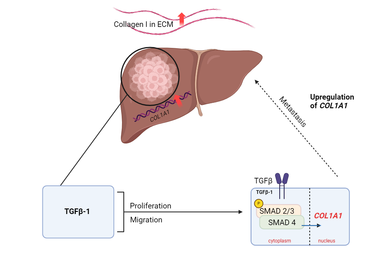
This study aim to determine how COL1A1 impacted the proliferation and migration behavior of Hepatocellular carcinoma and how TGF-β1 regulates the expression level of COL1A1
Hepatocellular Carcinoma (HCC), a major subset of liver
cancer, stands as a global health concern, accounting for over
700,000 annual deaths annually. Current treatments offer limited
prognostic efficacy, leading to persistent complications and tumor
recurrence. A deeper comprehension of novel targets against HCC
progression is imperative due to ineffective clinical treatments.
Currently, many cell transduction pathways have been identified and
correlated to excess Collagen I production in HCC, as Collagen I is
an indicator of HCC progression; however, the involvement of
Transforming growth factor-β1 (TGF-β1), which plays an important
role in HCC development, and the gene encoding Collagen type I
(COL1A1) in HCC remains unclear. This study aims to elucidate the
previously unknown relationship of the TGFβ pathway and the COL1A1
gene in HCC by testing how TGF-β1 regulates the expression level of
COL1A1 and its impact on the proliferation and migration of
hepatocellular carcinoma cells. HepG2 cells were cultured and COL1A1
was knocked down via siRNA transfection to assess changes in
proliferation and migration rates of HCC, in vitro. These cells were
then treated with TGF-β1. Based on the findings, COL1A1 is
overexpressed in HCC. Upregulation of COL1A1 facilitates the
proliferation and migration of HCC cells through the TGFβ pathway.
The correlation of TGF-β1 and COL1A1 upregulation introduces a novel
therapeutic target against HCC, deterring HCC cell progression by
reducing ambient levels of collagen I in the carcinoma tissue.
The graphical abstract was created in BioRender (BioRender, n.d.).
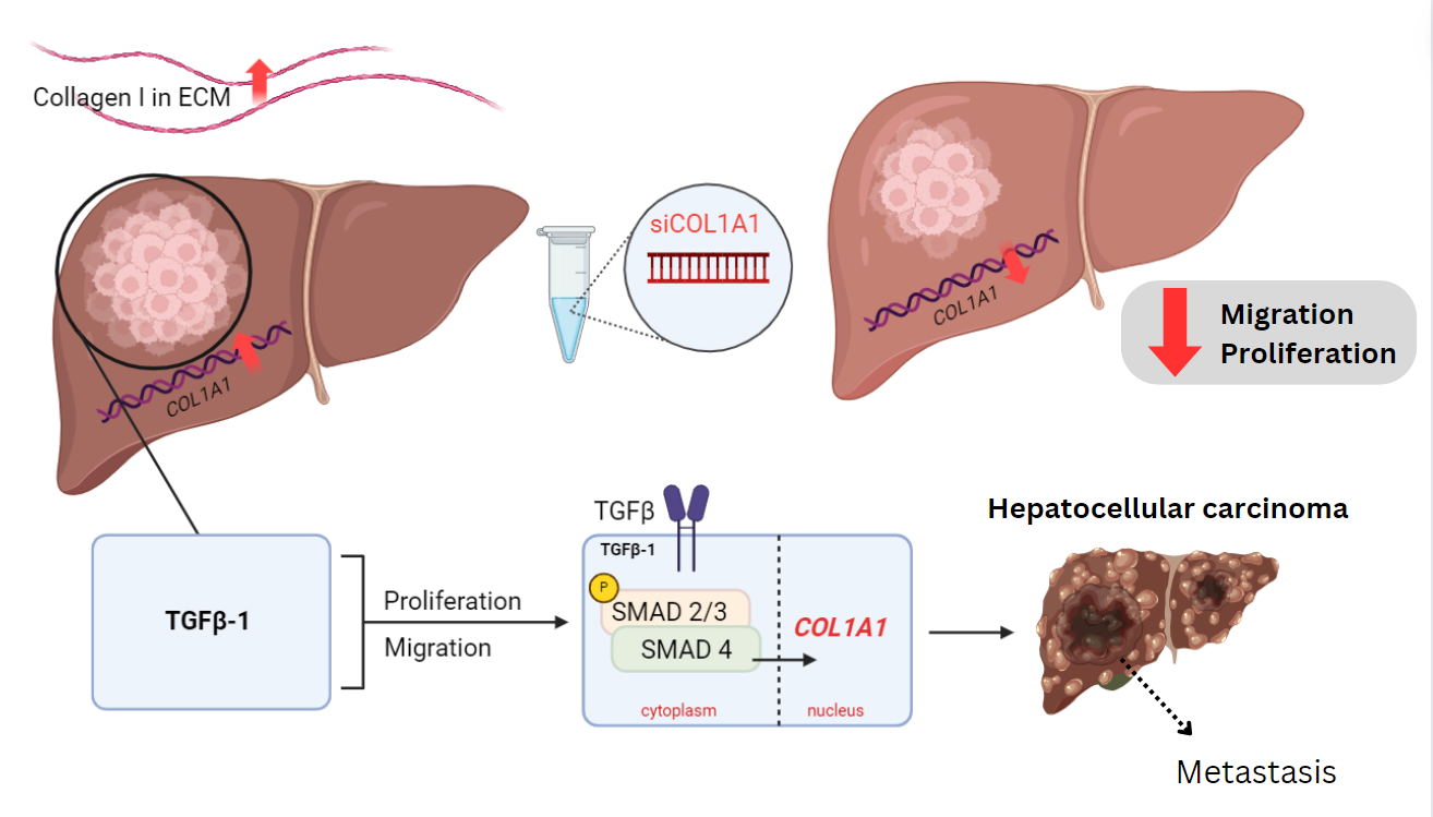
How does TGF-β1 regulate the expression level of COL1A1 and impact the proliferation and migration of hepatocellular carcinoma cells?
HepG2 cells treated with TGF-β1 will have increased COL1A1 expression, enhancing hepatocellular carcinoma cell proliferation and migration.
Liver cancer is one of the most malignant types of cancer, accounting for over 700,000 annual deaths worldwide (Ferlay et al., 2015). The most common form of liver cancer is Hepatocellular Carcinoma (HCC), which has emerged as the third leading cause of cancer-associated mortality worldwide. Currently, strategies against HCC include transplantation and target drugs; however, these treatment options lack proper prognosis, causing complications and tumor recurrence to remain a common occurrence. The lack of effective clinical treatments requires a further understanding of novel targets against hepatocellular carcinoma progression. Extracellular Matrix The extracellular matrix (ECM), a three dimensional structure with distinct biochemical and biomechanical properties, is an important component of the tumor microenvironment. Changes in the composition of the ECM have been shown to promote tumor growth in HCC (Khalfallah et al., 2011). Collagens are essential in ECM and can regulate tumor cell behavior (Wang et al., 2020). Collagen Type I is a major component of the liver ECM and is significantly upregulated in 83.7% of human liver cancer specimens compared with their adjacent non tumor tissue (Liu et al., 2021). Excess collagen in the tumor microenvironment creates a cycle of upregulation, further elevating levels of collagen I in the HCC microenvironment. The type I collagen network helps the basement membranes interact with nearby cells, playing a crucial role in cell migration and proliferation (Wang et al., 2020). The COL1A1 gene, alongside another alpha1 and alpha2 chain, provides instructions for making one component of type I collagen, which is upregulated in HCC as HCC progresses towards late stage carcinoma (Alomowitch et al., 2009). The upregulation of COL1A1 can generate a modified extracellular matrix environment that promotes the survival, proliferation, metastasis, and invasion of liver cancer cells (Wang et al., 2020). Therefore, the knockdown of the COL1A1 gene is a promising target to reduce HCC cell progression and moderate disease progression. TGF-β1 The process of HCC initiation, development, and progression involves various complex signaling pathways and networks, which are controlled by several oncogenes and tumor suppressor genes. Included in these pathways is the Transforming growth factor-β (TGF-β) signaling pathway, which plays important roles in several cellular processes, including proliferation and migration (Gonzalez-Sanchez et al., 2021). The TGF-β pathway’s involvement in cancer progression presents as an excellent therapeutic target. In the TGF-β pathway, Transforming growth factor-β1 (TGF-β1) binds to the TGF-β1 receptor, which induces the phosphorylation of Smad2/3 proteins. Phosphorylated Smad2/3 form a complex with Smad4, which translocates to the nucleus and regulates gene transcription through the interaction with various transcription factors (Ezzoukhry et al., 2016). TGF-β1 and Smad proteins are highly expressed in HCC cell lines and tissues, and play a significant role in the transcription of several genes, including COL1A1 (Shi et al., 2020). It has been identified that TGF-β1 stimulates the expression of COL1A1 and COL1A2 genes at the transcriptional and protein level; however little is known about TGF-β1’s relationship with COL1A1 and collagen I upregulation with respect to specific regulatory mechanisms relevant to HCC (Cutroneo et al., 2007). This study focuses on the role TGF-β1 plays in COL1A1 upregulation in HCC via the SMAD pathway.
Equipment and Materials
All materials required
for this project were provided and paid for by Dr. Mullen’s Research
Laboratory in the Department of Medicine/Division of
Gastroenterology at UMass Chan Medical School. HepG2 cells were
purchased from the American Type Culture Collection (ATCC) (HEPG2 -
HB-8065 | ATCC, n.d.). The HepG2 cells were cultured in Dulbecco's
Modified Eagle Medium (DMEM), supplemented with GlutaMAX™, 10% Fetal
Bovine Serum (FBS), and 1% Penicillin-Streptomycin, purchased from
ThermoFisher. 1X Phosphate Buffered Saline (PBS), Dulbecco's
Phosphate Buffered Saline (DPBS), and 0.25% Trypsin-EDTA, phenol red
were purchased from ThermoFisher. Opti-MEM™ was also purchased from
ThermoFisher. Dharmafect-1 was purchased from Fisher Scientific.
SiRNA smart pool against human COL1A1 and non-targeting control #5
were purchased from Dharmacon (Horizon Discovery Group plc, Level
Biotechnology, Inc, New Taipei City, Taiwan). MTT Cell Viability
Assay was purchased from Thermo Fisher, and data was collected using
a plate absorbance reader. Human TGF-beta 1 Recombinant Protein and
COL1A1 antibody were purchased from Thermo Fisher. A nanometer, cell
counter, and imaging software was also utilized to collect results.
In Silico Analysis of Cancer Expression
COL1A1 and TGF-β1 gene
expression profiling and correlative studies were performed using
the Gene Expression Omnibus (GEO) human hepatocellular carcinoma
microarray dataset and The Cancer Genome Atlas (TCGA) liver cancer
hepatocellular carcinoma (LIHC) cohort. Data from TCGA was analyzed
using the TCGA portal using the University of California Santa Cruz
(UCSC) Xena functional genomics explorer (Ma et al., 2019).
Cell Culture
The HepG2 cells were cultured at 37°C, in a 5% humidified
carbon dioxide incubator. Cells were cultured in media composed of
DMEM, 10% FBS, and 1% penicillin-streptomycin, and were grown for 48
hours in a 10 cm culture dish. Media was changed daily, and cells
were split weekly in accordance with assays.
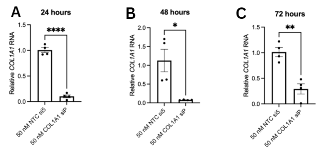
Testing COL1A1 siRNA knockdown Efficiency
Prior to conducting the proliferation and
migration assays, the knockdown efficiency of COL1A1 siRNA was
confirmed. Reverse siRNA transfection was utilized by plating COL1A1
siRNA and Non-targeting control (NTC) siRNA in 12-well plates. NTC
and COL1A1 siRNA were mixed with 6 μL of Dharmafect-1 and 1188 μL of
optimem. 478.8 μL of siRNA/transfection solution composed of 50 μM
NTC5 siRNA, DPBS, and Optimem was added to each well of a 12-well
dish. HepG2 cells were transferred and resuspended in 8640 μL of 16%
FBS. 720 μL of HepG2 cells were added to each well, and cells were
incubated in siRNA/transfection solution for 72 hours at 37°C before
harvesting. After treatment, cells were harvested, and total RNA was
extracted using TRIzol reagent. After following harvesting
protocols, mRNA levels of COL1A1 were measured using a quantitative
real-time polymerase chain reaction (qRT-PCR).
Cell Viability Assay
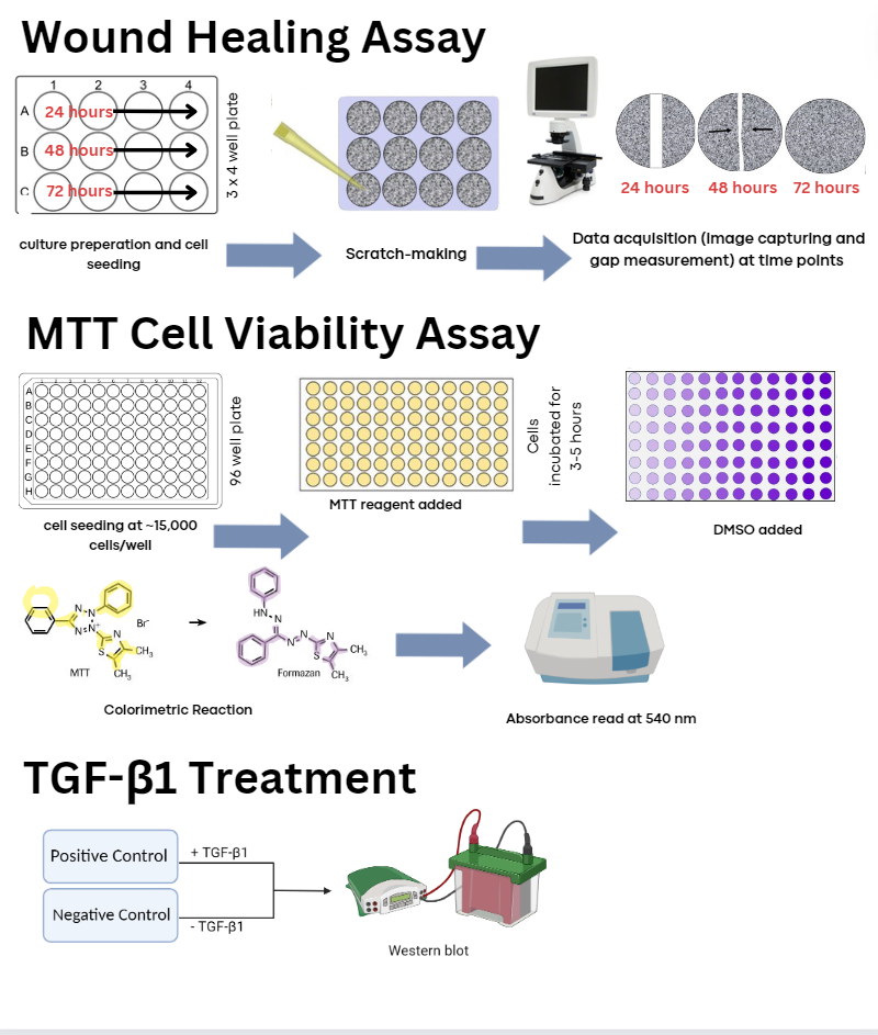
The MTT solution was prepared by mixing MTT with culture media.
HepG2 cells were seeded in a medium containing 10% FBS 24 hours
before performance of assay. 50 µL of serum-free media and 50 µL of
MTT solution was added into each well. The plate was incubated at
37°C for 3 hours, and after incubation 150 µL of MTT solvent was
added into each well. Absorption was read at OD=540 within 1 hour of
added solvent. Cell proliferation analysis was conducted by
averaging the duplicate reading for each sample and subtracting the
culture medium background from the assay reading to get the correct
absorbance proportional to cell number. siRNA knockdown efficacy was
quantified using qtPCR.
Wound Healing Migration Assay
Cells were seeded in 6-well plates with medium containing 10% FBS, then
cultured to 95–100% confluence. A scratch along the center of the
confluent adherent cells layer was made with a sterile p10 pipette
tip. Cell migration images were captured at 0, 12, 24 and 48 hours
after the scratch was made under a microscope and analyzed using
microscopy imaging analysis.
Western Blotting Analysis
Cellular protein lysates were isolated using a Protein Extraction Kit. An
equal amount of protein lysate sample was loaded in each lane and
subjected to sodium dodecyl sulfate polyacrylamide gel
electrophoresis (SDS-PAGE) along with a molecular weight marker
following manufacturers instructions. Relative band intensity was
analyzed using a Bio-Rad image software.
Statistical Tests
All assays were performed in triplicate. Comparisons between control and
target groups with normal distribution between the groups was tested
using a t-test or one-way ANOVA test. Comparison of means without a
normal distribution between the groups was done using Mann-Whitney
U-test or Kruskal-Wallis nonparametric tests. A p-value of < 0.05 was
considered statistically significant.
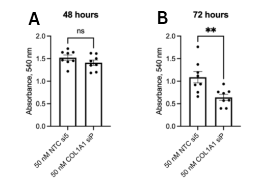
In this study, we investigated the role of COL1A1 in hepatocellular carcinoma tumorigenesis, as well as the involvement of TGF-β1 in COL1A1 expression levels. COL1A1 and type I collagen have been found to be upregulated in HCC samples compared to normal liver samples, conveying Type I collagen is an inducer of HCC (Zheng et al., 2017). Expression levels of TGF-β1 and COL1A1 in HCC were validated by screening the expression patterns of upregulated genes in liver cancer from the TCGA-LIHC database. Based on the upregulation of both genes in HCC samples, COL1A1 and TGF-β1 promote the growth of HCC cells. This study finds that TGF-β1 promotes HCC tumorigenesis by upregulating COL1A1. Knocking down COL1A1 via siRNA proved to be a potent inhibitor of the COL1A1 gene, decreasing mRNA expression of COL1A1 by 5 folds compared to the NTC. The confirmation that siCOL1A1 was successful in its knockdown of COL1A1 validated the use of siCOL1A1 for further assays involved in assessing the role of COL1A1. To further understand the role of COL1A1 in HCC, we determined the migration ability of cells with knocked down COL1A1 expression, finding that the suppression of COL1A1 reduced the migration of the HepG2 cells. Reduced migration rates of the NTC group indicate that COL1A1 promotes tumorigenesis and metastasis. COL1A1 upregulation in HCC cells also indicates increased proliferation rates, conveying the inhibition of COL1A1 can reduce the progression of HCC. The effect of COL1A1 on the growth and survival of HepG2 cells was measured using the MTT assay. We found that siCOL1A1 stunted the proliferation of HepG2 cells, as indicated by the lower MTT signals obtained upon siRNA transfection in comparison to the untreated cells. siCOL1A1 is a potent inhibitor of cell proliferation presumably because it suppresses the overexpression of COL1A1 in liver cell lines, which is consistent with a study that found the inhibition of COL1A1 reduces protein supply and number of organelles, regulating the cell cycle progression (Xu-Huang Fu et al., 2019) Using HepG2 cultured cells, we confirmed that TGF-β1 stimulation increases the production of the COL1A1 gene. When cells were treated with TGF-β1, a western blot showed an increased expression of Collagen I, indicating that the treatment of TGF-β1 upregulated COL1A1 expression. The relationship between TGF-β1 and COL1A1 upregulation indicates that TGF-β1 expression induces COL1A1 gene expression. This finding is validated by a study in which it was found that without the presence of type I collagen, TGF-β1 is unable to promote invadosome formation and associated matrix degradation (Ezzoukhry et al., 2016). Based on these results, it will be interesting to test the impact of TGF-β1 stimulation on HCC migration and proliferation rates in order to further validate the findings of this study. It is also important to test the knockdown of the SMAD pathways against the COL1A1 gene to further optimize potential therapeutic targets. Future work also includes investigating the biochemical mechanics of the TGF-β1 and COL1A1 relationship by exploring natural inhibitors.
The findings of this study shed light on the relationship between COL1A1, TGF-β1, and the progression of hepatocellular carcinoma. Our findings support that COL1A1 promotes HepG2 migration and invasion by activating TGF-β1. By employing knockdown techniques targeting COL1A1, the study elucidated the role of COL1A1 in regulating cell proliferation and migration within HCC. The reduction in cell proliferation and migration following COL1A1 knockdown underscores its significance in driving the aggressive behavior of HCC cells and promoting tumorigenesis. The investigation into the involvement of TGFβ1 in COL1A1 upregulation provides valuable insights into potential signaling pathways underlying HCC progression. Through the treatment of cells with TGFβ1, the study uncovered a potential regulatory mechanism by which TGFβ1 promotes COL1A1 expression, thus implicating the TGFβ1/COL1A1 axis as a potential therapeutic target for HCC intervention. These findings not only deepen our understanding of the molecular mechanisms driving HCC progression but also offer promising avenues for the development of targeted therapeutic strategies aimed at inhibiting COL1A1-mediated pathways in HCC.