STEM is a course taught by Dr. Crowthers. The first part of the year in this course is spent in STEM I, which is our independent research project. During this, you learn all the necessary skills to become a researcher through conducting your own research project on a topic of your choosing. For STEM I, I chose to investigate the effects of polyethylene and polypropylene microplastics on endothelial cells and their functionality.
This project aims to discover the effects of microplastics on human health as that is a field that there is minimal research in. This study utilizes three concentrations of polyethylene and polypropylene microplastics to investigate their effects on endothelial cells. Through conducting a trypan blue exclusion assay and image analysis, this study determined the effects on both cell morphology and viability. It was determined that both types of microplastics do indeed cause decreased cell viability and great changes to cell morphology. This creates the need to investigate further into the effects of microplastics and develop a way to extract them from the human body because if not done, the presence of these microplastics could have devastating effects on human health.
Endothelial cells are a vital group of cells found in both blood and lymphatic vessels and are responsible for many physiological functions in the human body, the most significant being production of the glycocalyx. The glycocalyx is a carbohydrate-rich fibrous layer that lines the surface of endothelial cells and plays several key roles in the human body. However, small plastic particles, known as microplastics, can be found in the blood vessels, potentially posing a threat to the endothelial cells and the glycocalyx. These microplastics enter the body through various methods, including inhalation and ingestion. They are newly discovered and thus there is little research on them – but they are known to be very toxic.The goal of this study is to investigate the effects these microplastics can have on endothelial cells, and consequently, the glycocalyx. This study utilizes two of the most common types of microplastics - polyethylene and polypropylene - to investigate the effects of microplastics on the viability of human lung endothelial cells. A trypan blue exclusion assay, ImageJ, and Cell Profiler were used to analyze cell viability and morphology. As shown through the study, microplastics do impact both cell viability and morphology. This study aims to showcase the dangers of microplastics in hopes of conveying the true threat these small plastic particles pose. Through this, the study hopes to show the need for further research into microplastics and for an alternative form of plastic.
How do microplastics of various types affect cell viability and morphology of endothelial cells?
It is hypothesized that the higher the concentration of microplastics, the lower the cell viability and the greater the changes to cell morphology. Additionally, polyethylene microplastics will yield, overall, lower cell viability and greater changes to cell morphology.
Microplastics are plastic particles that can range from 1 nanometer to 5 millimeters in size. These can be found everywhere: in the air we breathe to the water we drink. Even though they are so prevalent, microplastics were first discovered in 2004 by Marine Biologist Richard Thompson (Schmid et al., 2021). However, microplastics weren’t detected in the human body until 2022 when researchers in the Netherlands found microplastics in human blood samples (Roeloffs, 2024). Microplastics can be made of various materials and even though little is known about them, existing research about them shows their harmful effects on other living organisms. This raises the issue of the lack of knowledge regarding their effect on humans and thus the dangers that they are constantly exposed to.
Types of Microplastics
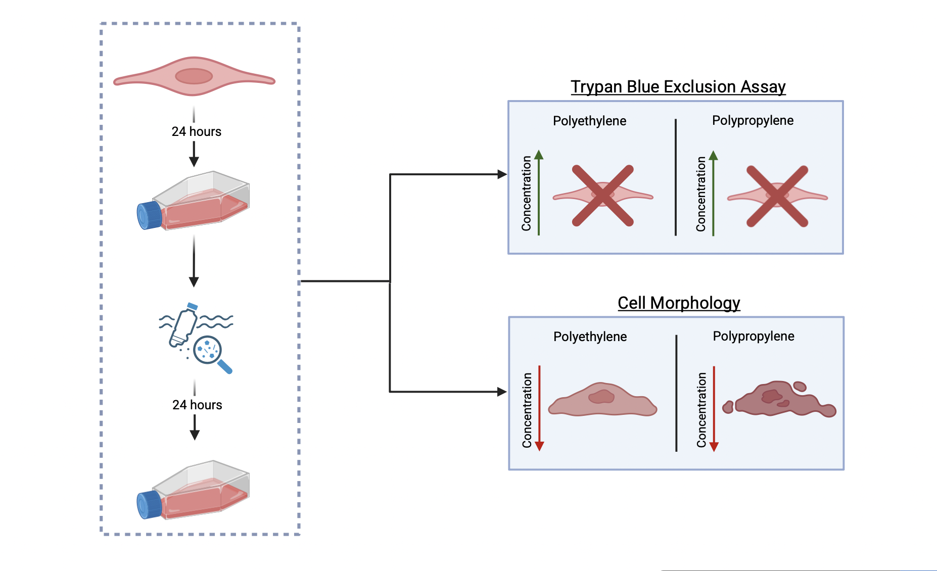
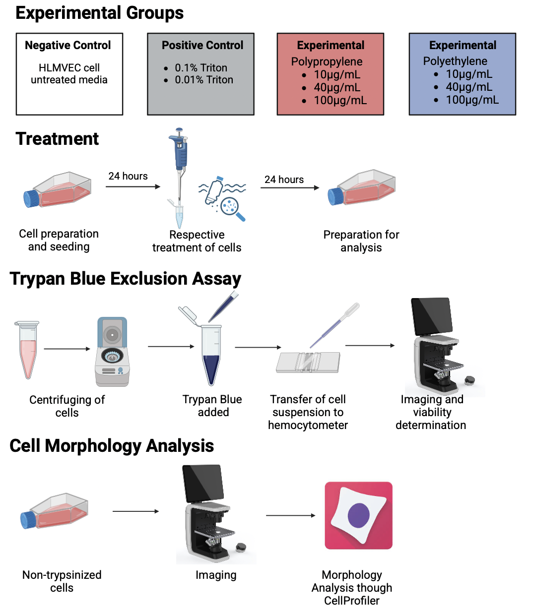
There are two main categories of microplastics: Primary and Secondary, which are differentiated by how they were created. Primary microplastics are produced by manufacturers to be small. They can come in the form of either pellets (called nurdles) or in the form of microbeads, which are often manufactured for the cosmetic and pharmaceutical industry (Microplastics – Pollution Tracker, 2016). Secondary microplastics, on the other hand, result from the breakdown of larger plastics – often beach litter or laundry (Microplastics – Pollution Tracker, 2016). Overall, there are 13 types of microplastics. However, the most common types of microplastics are polyethylene, polypropylene, and polystyrene (Olabode & Fulmer, n.d.).
Effects of Microplastics
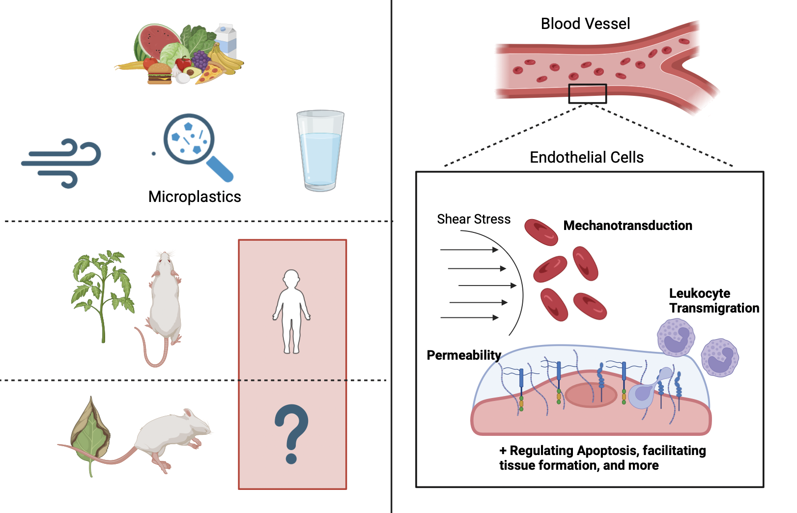
Microplastics are a new discovery, and thus little research has been done on them. However, studies have shown that microplastics are extremely toxic, inducing oxidative damage, DNA damage, organ dysfunction, metabolic disorders, weakened immune responses, neurotoxicity, reproductive toxicity, and developmental toxicity (Li et al., 2023). However, most of the studies done have focused on organs directly exposed to microplastics, such as the lungs and small intestines. Minimal studies have been done on indirectly exposed organs, such as the heart, and correlating parts, such as blood vessels.
Endothelial Cells
Endothelial cells line all blood and lymphatic vessels found in the human body and thus play a key role in blood transport and human survival. In fact, these cells are often the first line of defense for any ingested toxin prior to entering the body. Endothelial cells are found most commonly in arteries, veins, capillaries, and lymph capillaries. In lymph capillaries, endothelial cells function as a semi-permeable barrier, allowing for the transport for lymph fluid along with facilitating the movement of immune cells to lymph nodes – which is necessary for the immune system to function (Cleveland Clinic, 2022). In arteries, veins, and capillaries, endothelial cells are responsible for producing glycocalyx, a vital substance for human survival (Villalba et al., 2021).
Vascular Endothelial Glycocalyx
The endothelial glycocalyx is a carbohydrate-rich, fibrous layer that lines the luminal surface of endothelial cells and blood vessels, thus playing many vital roles in human survival (Reitsma et al., 2007). One of the functions of the glycocalyx is permeability. The glycocalyx regulates transport of water proteins, and other essential molecules from the blood to the outside of blood vessels. Along with this, it can restrict certain molecules from passing through endothelial cells, thus protecting blood vessels. This restriction can be attributed to the structure and negative charge of the glycocalyx (Jin et al., 2021).The glycocalyx is also necessary for inflammation. It plays an essential role in the occurrence and development of inflammations, which is a defense mechanism that allows the body to fight off infections and heal. When inflammation occurs, the glycocalyx is shed, which makes it easier for leukocytes to bind to endothelial cells. This binding is essential for immune response. The glycocalyx plays a key role in the anticoagulant process as well. Under certain conditions, the interaction between endothelial cells and blood cells can be altered, avoiding thrombosis. By interacting with antithrombin III, thrombomodulin, tissue factor pathway inhibitors, and other molecules, the glycocalyx can induce anticoagulant effects (Jin et al., 2021). The glycocalyx is also necessary for sending signals. It detects changes in blood flow and then transmits that information to the endothelial cells. This enables the cells to respond with various morphological responses. The glycocalyx can also regulate apoptosis of the endothelial cells, allowing them to die whenever necessary and keeping them alive otherwise. A key function of the glycocalyx is cerebrovascular micro-homeostasis. The glycocalyx is responsible for maintaining the barrier function of the cerebral blood vessels. It regulates the permeability of the blood-brain barrier (BBB) and plays a key role in cerebrovascular coagulation and neuroinflammatory processes (Jin et al., 2021).
Equipment and Materials
All materials used, except microplastics, were purchased and provided by the Dr. Mensah Lab at Worcester Polytechnic Institute. The cells used in the project were Human Lung Microvascular Endothelial Cells (HLMVEC) that were cryopreserved in basal medium with 10% FBS and 10% DMSO purchased from CellApplications. The growth medium used for the HLMVEC cells was a specialized Human Microvascular Endothelial Cell media developed for HLMVEC cells purchased from CellApplications. For the positive control, 10% Aqueous Triton X-100 Detergent Surfact-Amps Solution purchased from ThermoFisher was used. Polyethylene and propylene wax microplastics were used for experimental groups. To create the various concentrations of microplastics, Dimethyl sulfoxide (DMSO) purchased from Sigma Aldrich was used. Phosphate buffered saline (PBS) and trypsin purchased from Sigma Aldrich were also used. The cells were stored in an incubator kept at a constant 37° Celsius and 5% Carbon Dioxide once thawed. For analysis, a hemocytometer and 0.4% Trypan Blue Solution purchased from ThermoFisher was used.
Creation of Dosage Concentrations
For this study, three concentrations of two different microplastics were chosen based off a previous study conducted by Wang et al., 2022. Various dilutions were used to create the three concentrations of each type of microplastic. The starting dilution was 0.01 grams of microplastic in 1 mL DMSO. For the 10ug/mL, 5uL of the starting solution was added to 5mL of media. For 40ug/mL, 20uL of the starting solution was added to 5mL of media. For 100ug/mL, 50uL of the starting solution was added to 5mL of media.
Cellular Treatment
This technique entitled dividing 1 million cells, right after thawing, into eight T-25 flasks, which entailed 125,000 cells per flask. Cells were incubated for 24 hours to allow sufficient cell growth and were then treated with their respective dosages. Cells were incubated for another 24 hours with their treatments, based on a collection of previous studies, and imaged for later analysis. Then, cells were trypsinized and stained using 0.4% Trypan Blue Solution to be analyzed using a hemocytometer.
Morphology Determination
Cell Profiler was used to analyze the morphology of the cells. The specific pipeline used was as follows: (1) Rescale Intensity – Divide by the image’s maximum, (2) Enhanced Edges – Sobel in all directions, (3) Identify Primary Objects – Range from 40-200, Adaptive, Otsu, 3 classes, and (4) Measure Object Size and Shape – Area, Perimeter, Eccentricity, and Maximum Radius
Welch’s T-Test
Welch’s T-Test was used to analyze cell morphology data. A t-test was chosen as it is the most efficient way to calculate significance between two variables and for the purposes of this project, was deemed necessary. Welch’s corrections were applied before conducting the t-test as each group had different standard deviations and group sizes.
Figure 1: Area of Endothelial Cells represented through (a) pixel intensity and (b) fold change
Figure 2: Perimeter of Endothelial Cells represented through (a) pixel intensity and (b) fold change
Figure 3: Maximum Radius of Endothelial Cells represented through (a) pixel intensity and (b) fold change
Figure 4: Eccentricity of Endothelial Cells represented through (a) pixel intensity and (b) fold change
Not able to view the PDF? Click here
Graphical Abstract, Background, and Procedure were created through BioRender (BioRender, n.d.).